3 types of Blood Vessels in the body
Blood Vessels
These are hollow tubular vessels in which the blood flows.
what are blood vessels?
Blood is transported throughout the body through blood vessels, which are essential parts of the circulatory system. Arterioles, veins, and capillaries are the three primary categories.
Table of Contents

Blood arteries are essential for controlling blood flow, keeping blood pressure stable, and delivering nutrients and oxygen to tissues. They also aid in the body’s elimination of waste. The complex system of blood arteries ensures that the circulatory system operates properly, promoting general health and well-being.
There are three types of Blood Vessels.
Arteries
These are thick-walled and deeply located blood vessels, which always carry the blood away from the heart to various, body parts. these have narrow lumens but do not have valves. in them, the blood flows at high pressure and high speed. these generally carry oxygenated blood.
what are arteries?
Arteries are blood arteries with strong walls that transport oxygenated blood from the heart to other organs and tissues. They feature a thick, elastic covering that controls blood flow and helps keep blood pressure stable.
A specific form of blood channel called an artery transports oxygenated blood from the heart to the body’s many organs and tissues. They can endure the enormous pressure produced by the heart during systole (contraction) thanks to their thick walls and robust, elastic construction.
How many layers of arteries?
The interior endothelium, the middle layer of smooth muscle, and the outside layer of connective tissue make up the three layers that make up the walls of arteries. Atherosclerosis gives arteries the ability to control blood flow and maintain blood pressure.
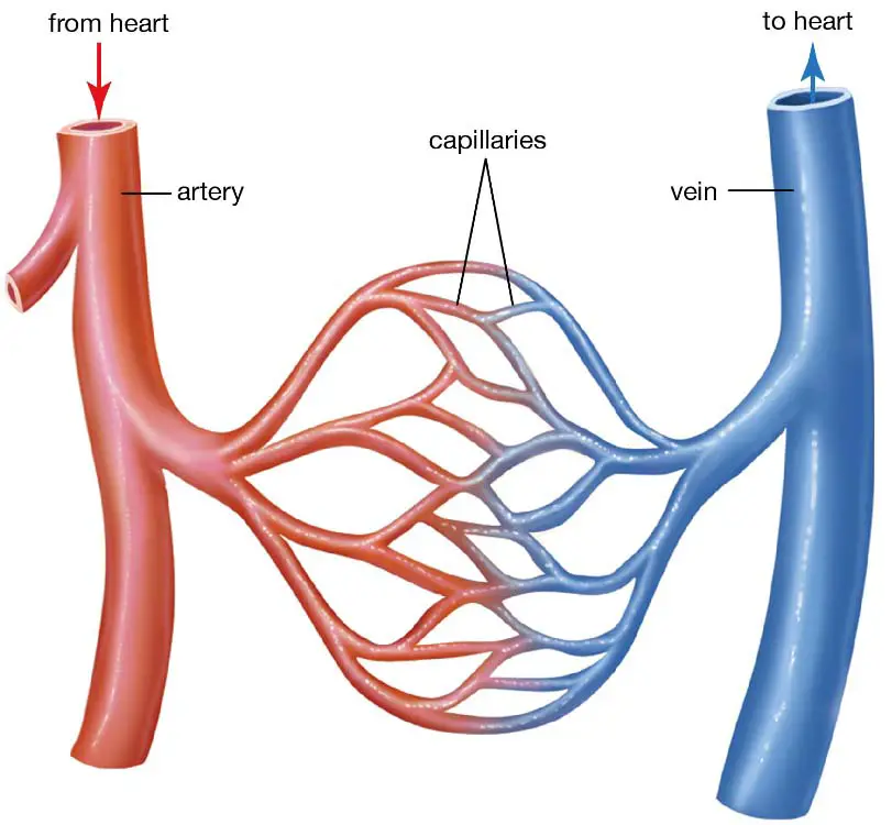
Arterioles, which are even smaller vessels than arteries, split off from arteries to form tiny capillaries. These capillaries let the cells and tissues in the area exchange waste, nutrients, and oxygen.
Compared to veins, arteries are usually located deeper within the body. They also contain a pulse, which is felt as a rhythmic throbbing feeling. They are essential for providing organs and tissues with oxygen-rich blood, ensuring their healthy function.
Veins
These are thin-walled and superficially located blood vessels, which always carry blood from various, body parts generally to the heart. these have wider lumens and valves in them to prevent the backflow of blood. in them, blood flows at low pressure and low speed.
Conversely, veins return blood that has lost oxygen to the heart. They have included valves and thinner walls to stop backflow. Blood flow via veins is aided by the surrounding muscles contracting.
what are veins?
Blood channels called veins are responsible for returning tissues and organs’ deoxygenated blood to the heart. They assist arteries in ensuring appropriate blood flow throughout the body.
Compared veins to arteries?
Compared to arteries, veins have thinner walls and valves, which assist stop blood from flowing backwards. Particularly in the lower extremities, these valves provide effective blood flow in the face of gravity. Venous blood flow is aided by the surrounding muscles contracting as people move.
Veins are not throbbing like arteries and are frequently found nearer the surface of the body. They create a complex network throughout the body and are more numerous than arteries.
What is superior vena cava?
The superior vena cava, which gets blood from the upper body, and the inferior vena cava, which receives blood from the lower body, are the two primary veins that develop after receiving blood from the capillaries and progressively combine into bigger vessels. These significant veins return oxygen-depleted blood to the heart for circulation.
Veins are essential for the circulatory system’s smooth operation and for bringing blood back to the heart.
Capillaries
These are the thinnest blood vessels. and formed by division and re-division of arteriole inside the body organs. each is lined by a single layer of flat cells called the endothelium, which allows the exchange of materials between blood and the body cells through tissue fluid.
what are capillaries?
The tiniest and most abundant blood veins are capillaries. They link arteries and veins, allowing for the exchange of waste materials, nutrients, and oxygen with neighbouring tissues. Since capillary walls are so tiny, diffusion is effectively possible.
The tiniest and thinnest blood vessels in the body, capillaries join veins and arteries. Their main job is to help the blood and surrounding tissues exchange nutrients, hormones, waste products, and oxygen.
Capillaries can access practically every cell in the body because to their minuscule diameter. Because of the single layer of endothelial cells that makes up their very thin walls, things diffuse effectively.
what are the functions of capillaries?
Carbon dioxide and waste materials are eliminated while oxygen and nutrients are given to tissues through the capillary exchange mechanism. The capillaries’ thin walls allow for this exchange to happen, which is fueled by pressure and concentration gradients.
Additionally, capillaries are essential for controlling blood flow. To provide an adequate supply depending on metabolic needs, they can dilate or constrict to control the volume of blood reaching particular tissues or organs.
The wide capillary network offers a large surface area for exchange, facilitating the delivery of necessary chemicals and the elimination of waste items, and therefore enhancing the body’s general health and functionality.

Structure of Blood Vessels
What is the Structure of Blood Vessels?
A blood vessel ( artery or vein) is formed of three layers., these are tunica Interna tunica media and tunica external.
Tunica Interna is the innermost layer and is formed of the inner endothelium of flat cells and the outer elastic membrane of yellow fibrous tissue.
Tunica media is the middle layer and is formed of circular smooth muscle fibres and elastic yellow fibres.
Tunica Externa is the outermost layer and is formed of collagen-rich areolar connective tissue.
What are Interna, media and Externa Tunica?
Blood vessels are made up of three primary layers, or tunics, that provide strength, flexibility, and blood flow regulation.
- The Tunica intima is the layer that is closest to the blood. It is made up of an extremely thin layer of endothelial cells, which have a smooth surface to reduce friction and support blood flow. A subendothelial layer is also present in the tunica intima and serves as support.
- Tunica media: Elastic and smooth muscle fibres make up this layer’s middle layer. The smooth muscle permits the blood vessel to be vasoconstrictive (narrowed) and vasodilated (widen), controlling blood flow and blood pressure. Because of the elasticity provided by the elastic fibres, the vessel can expand and recoil.
- Adventitia (tunica externa): Connective tissue makes up this skin’s outer layer. It gives the blood vessel structural stability and defence. Additionally, it has nerve fibres and tiny blood vessels called vasa vasorum that feed the bigger blood artery with nutrients and oxygen.
Depending on the kind of blood artery, these layers’ configuration and thickness might differ. Veins have thinner walls with valves to stop backflow, whereas arteries have a larger tunica media. Capillaries, which facilitate the flow of chemicals between blood and tissues, only have a single layer of endothelial cells.
Difference Between Artery and Vein
What is the difference between artery and vein?
| Artery | Vein |
|---|---|
| 1) Endothelial cells of tunica interna are more flattened. | 1) Endothelial cells of tunica interna are less flattened. |
| 2) The elastic membrane of Tunica interna is more developed so is more elastic. | 2) The elastic membrane of Tunica interna is less developed so is less elastic. |
| 3) Tunica media is better developed so is more contractile. | 3) Tunica media is less developed so is less contractile. |
| 4) Tunica externa is less developed so is less strong. | 4) Tunica externa is more developed so is more strong. |
| 5) Lumen is small. | 5) Lumen is large. |


